X
X
Leaving Community
Are you sure you want to leave this community? Leaving the community will revoke any permissions you have been granted in this community.
No
Yes
X
Inside NIF: The Cell: An Image Library
Inside NIF: The Cell: An Image Library
 The Cell: An Image Library™ is a open, easy to use repository of high-quality images, videos and animations of cells. Given their efforts to semantically annotate uploaded images, non-scientist users can quickly browse to a particular type of cell, or cellular process with ease.
NIF recently hosted a webinar with David Orloff, manager of CIL, who gave an excellent overview of their philosophy, technologies and future directions. It also inspired the NIF team to browse around in CIL, bringing to you some of the best neuroscience related images we could find - formatted in iPhone wallpaper size. Check them out below:
Image 1: Cultured hippocampal neurons, immunostained for MAP2 & PSD95

(Author: Dieter Brandner & Ginger Withers, Whitman College
Image 2: Labeled mitochondria in fibroblasts.

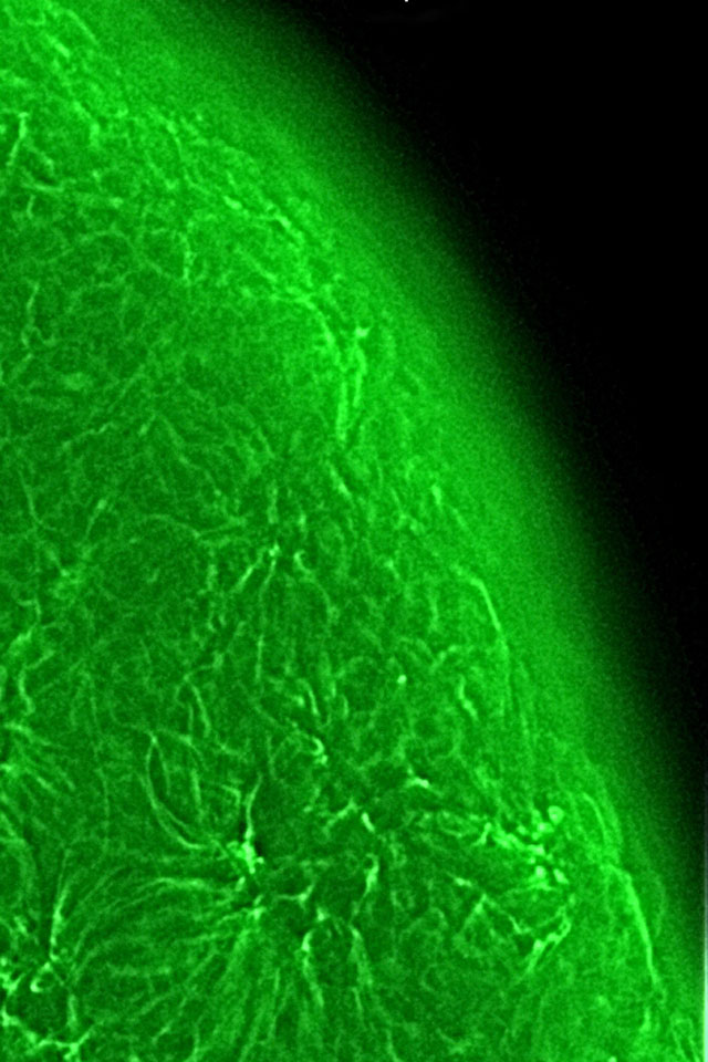
Image 3: Mouse Ovary with tublin labeled - NOT a planet!

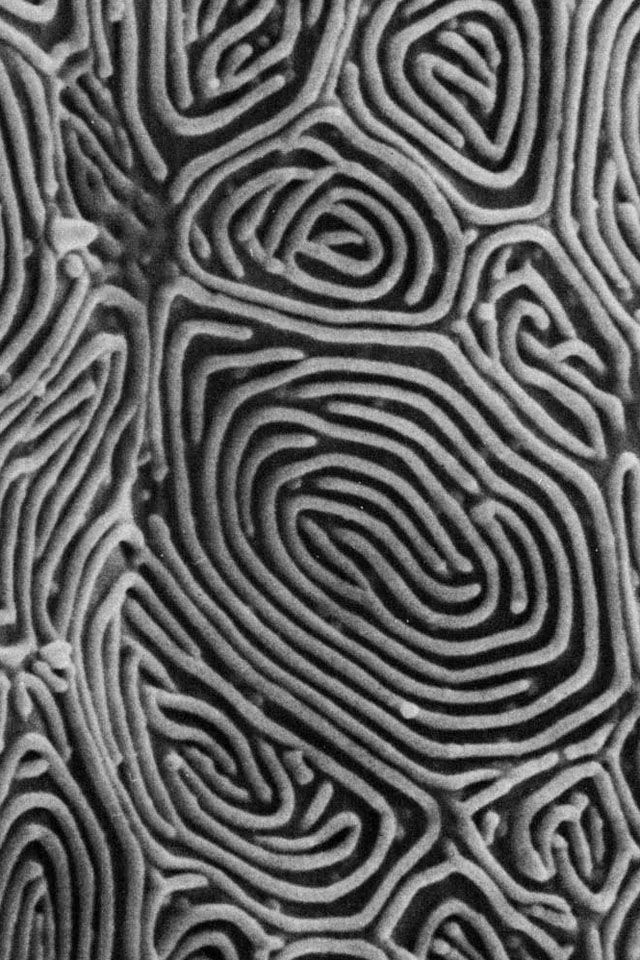
Image 4: Surface of epidermal cell

(Authors: Don W. Fawcett & Wolf Fahrenbach
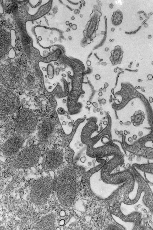
Image 5: Somatic basal bodies at cytoproct ridge

X


